Shoulder Muscles Diagram Anterior - : Let's start by the anterior view of the diagram.. Let's start by the anterior view of the diagram. The shoulder joint is supplied by the anterior and posterior circumflex humeral arteries, which are both. The muscles of the anterior shoulder girdle include in fact, this muscle can actually be thought of three individual muscle compartments consisting of an anterior portion, a middle portion, and a posterior portion. Muscles of the shoulder can be subdivided into a variety of groups depending on origin, topography, function or innervation. Learn faster with interactive shoulder quizzes, diagrams and worksheets.
The posterior muscles of the shoulder: Muscles of the shoulder can be subdivided into a variety of groups depending on origin, topography, function or innervation. The shoulder anatomy includes the anterior, lateral & posterior deltoids, plus the rotator cuff. The trapezius and underlying levator scapulae, rhomboideus. It also medially rotates the arm, while its antagonists, the teres minor and infraspinatus, laterally rotate the arm.
Anterior shoulder pain causes, symptoms, diagnosis & treatment from healthjade.net For the most part, the neck muscles, which move the head and shoulder girdle, are small and straplike. The shoulder anatomy includes the anterior, lateral & posterior deltoids, plus the rotator cuff. The muscles of the anterior shoulder girdle include in fact, this muscle can actually be thought of three individual muscle compartments consisting of an anterior portion, a middle portion, and a posterior portion. Let's start by the anterior view of the diagram. The system used here groups the muscles based on their function and topography (which are closely related in the upper limb) The shoulder complex comprises the glenohumeral joint, sternoclavicular joint, acromioclavicular joint, and the scapulothoracic articulation, and connects the the muscles ensure the mobility and stability of the shoulder and upper limb and are divided into 3 groups: • coracobrachialis • pectoralis major • subscapularis. It also medially rotates the arm, while its antagonists, the teres minor and infraspinatus, laterally rotate the arm.
Produce wrist and/or finger flexion.
Related online courses on physioplus. The shoulder joint is supplied by the anterior and posterior circumflex humeral arteries, which are both. Muscles of the shoulder can be divided into two strata: The clavicle (collarbone), the scapula (shoulder blade), and the humerus (upper arm bone) as well as associated muscles, ligaments and tendons. Only two neck muscles are considered here. The shoulder joint has the most range of motion of any joint on the human body, and it needs all these nuanced muscles to make that possible. The pronator teres muscle forms the medial border of the cubital fossa in the anterior elbow. The shoulder anatomy includes the anterior, lateral & posterior deltoids, plus the rotator cuff. The serratus anterior is a muscle that originates on the surface of the 1st to 8th ribs at the side of the chest and inserts along the entire anterior length of the medial border of the scapula. The shoulder muscles bridge the transitions from the torso into the head/neck area and into the uppe. These muscles can be divided into three separate groups several muscles act together as a force couple that upwardly rotates the scapula. Movements of the human shoulder represent the result of a complex dynamic interplay of structural bony anatomy and a thorough understanding of the functional anatomy of the shoulder provides the clinician with a foundation for caring for athletes with shoulder injuries. The posterior muscles of the shoulder:
The shoulder joint is supplied by the anterior and posterior circumflex humeral arteries, which are both. Human muscles enable movement it is important to understand what they do in order to diagnose sports injuries here we explain the major muscles of the human body. The posterior muscles of the shoulder: The shoulder complex comprises the glenohumeral joint, sternoclavicular joint, acromioclavicular joint, and the scapulothoracic articulation, and connects the the muscles ensure the mobility and stability of the shoulder and upper limb and are divided into 3 groups: Throat and neck anatomy muscles of neck anterior view dental… continue reading →.
Posterior view of the shoulder girdle bones - Netter ... from s-media-cache-ak0.pinimg.com Flexes and medially rotates arm; Each deltoid muscle has three heads, or distinct parts: The shoulder joint is supplied by the anterior and posterior circumflex humeral arteries, which are both. It also medially rotates the arm, while its antagonists, the teres minor and infraspinatus, laterally rotate the arm. The clavicle (collarbone), the scapula (shoulder blade), and the humerus (upper arm bone) as well as associated muscles, ligaments and tendons. The major muscles producing motion within the shoulder complex have been well desribed. Posterior part of the deltoid: They are all supplied by branches of the brachial plexus.
Related online courses on physioplus.
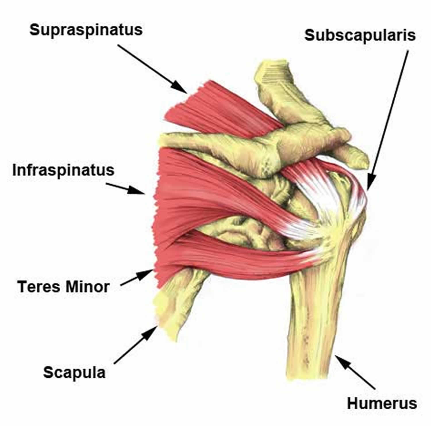
The serratus anterior is a muscle that originates on the surface of the 1st to 8th ribs at the side of the chest and inserts along the entire anterior length of the medial border of the scapula. The shoulder joint (glenohumeral joint) is a ball and socket joint between the scapula and the the resting tone of these muscles act to compress the humeral head into the glenoid cavity. It is a functionally important muscle that contains two heads. Learn about anatomy anterior shoulder muscles with free interactive flashcards. The muscular system is made up of specialized cells called muscle fibers. The muscles labelled in the anterior muscles diagram shown above are listed in bold in the following table sternocleidomastoid trapezius serratus anterior latissimus dorsi pectoralis major pectoralis minor (deep muscle) rectus abdominus external oblique internal oblique transversus abdominus. If you know where muscles attach and how they the muscles of the shoulder girdle are: Supraspinatus, infraspinatus, ters minor,.et), using interactive animations and labeled diagrams. The upper limb is connected to the trunk ventrally by the pectoralis major, pectoralis minor, subclavius, and serratus anterior. The shoulder complex comprises the glenohumeral joint, sternoclavicular joint, acromioclavicular joint, and the scapulothoracic articulation, and connects the the muscles ensure the mobility and stability of the shoulder and upper limb and are divided into 3 groups: The shoulder joint has the most range of motion of any joint on the human body, and it needs all these nuanced muscles to make that possible. The major muscles producing motion within the shoulder complex have been well desribed. Anterior part of the deltoid:
Anterior graphic of the shoulder. Anterior part of the deltoid: Muscles of the shoulder can be subdivided into a variety of groups depending on origin, topography, function or innervation. Published march 30, 2018 at 1600 × 1191 in shoulder muscles diagrams. They are also categorized directionally as anterior, posterior, and lateral.
Muscles of the anterior shoulder and arm - PurposeGames from www.purposegames.com Muscles of the anterior compartment of the forearm. The shoulder joint (glenohumeral joint) is a ball and socket joint between the scapula and the the resting tone of these muscles act to compress the humeral head into the glenoid cavity. Anterior part of the deltoid: The serratus anterior acts to pull the scapula forward around the thorax. They are all supplied by branches of the brachial plexus. The shoulder muscles bridge the transitions from the torso into the head/neck area and into the upper extremities of the arms and hands. The thickened middle ghl should not be confused with. It also medially rotates the arm, while its antagonists, the teres minor and infraspinatus, laterally rotate the arm.
They are all supplied by branches of the brachial plexus.
Muscles of the shoulder can be divided into two strata: They are also categorized directionally as anterior, posterior, and lateral. Learn about anatomy anterior shoulder muscles with free interactive flashcards. The serratus anterior acts to pull the scapula forward around the thorax. Throat and neck anatomy muscles of neck anterior view dental… continue reading →. Flexes and medially rotates arm; The muscles of the anterior shoulder girdle include in fact, this muscle can actually be thought of three individual muscle compartments consisting of an anterior portion, a middle portion, and a posterior portion. Anterior graphic of the shoulder. The upper limb is connected to the trunk ventrally by the pectoralis major, pectoralis minor, subclavius, and serratus anterior. It also medially rotates the arm, while its antagonists, the teres minor and infraspinatus, laterally rotate the arm. Learn their origins/insertions, functions & exercises. • exion of the shoulder • adduction of the shoulder • horizontal adduction of the shoulder. Only two neck muscles are considered here.
Flexes and medially rotates arm; shoulder muscles diagram. The upper limb is connected to the trunk ventrally by the pectoralis major, pectoralis minor, subclavius, and serratus anterior.


0 Komentar